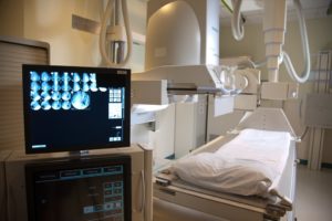
 How Are The X-Rays Made
X-rays are a form of radiation like light or radio waves. X-rays pass through most objects, including the body. Once it is carefully aimed at the part of the body being examined, an x-ray machine produces a small burst of radiation that passes through the body, recording an image on photographic film or a special detector.
Patient Expectation
The technologist will bring the patient into the room. Dependent on the area of interest, the patient may be placed on the x-ray table or the exam may be done standing. The x-ray beam will then be focused on the area of interest. Various x-rays may be taken at different angles to better visualize the area.
The length of the exam depends on the type of study that is ordered. Most general exams take just a few minutes.
What Are The Benefits And Risks?
Benefits
- No radiation remains in a patient’s body after an x-ray examination.
- X-rays usually have no side effects in the typical diagnostic range for this exam.
- X-ray equipment is relatively inexpensive and widely available in emergency rooms, physician offices, ambulatory care centers, nursing homes and other locations, making it convenient for both patients and physicians.
- Because x-ray imagine is fast and easy, it is particularly useful in emergency diagnosis and treatment
Risks
- There is always a slight chance of cancer from excessive exposure to radiation. However, the benefit of an accurate diagnosis far outweighs the risk.
- The effective radiation dose for this procedure varies.
- Women should always inform their physician or x-ray technologist if there is any possibility that they are pregnant.
A Word About Minimizing Radiation Exposure
Special care is taken during x-ray examinations to use the lowest radiation dose possible while producing the best images for evaluation. National and international radiology protection organizations continually review and update the technique standards used by radiology professionals.
Who Interprets the Results and How Do I Get Them?
A radiologist, a physician specifically trained to supervise and interpret radiology examinations, will analyze the images and send a signed report to your primary care or referring physician, who will discuss the results with you.
The results of a x-ray can be available almost immediately for review by your physician.
Follow-up examinations may be necessary, and your doctor will explain the exact reason why another exam is requested. Sometimes a follow-up exam is done because a suspicious or questionable finding needs clarification with additional views or a special imaging technique. A follow-up examination may also be necessary so that any change in a known abnormality can be monitored over time. Follow-up examinations are sometimes the best way to see if treatment is working or if an abnormality is stable or changed over time.
Fluoroscopy
Fluoroscopy is the procedure performed by a radiologist to view tissues and organs in motion. During the procedure, a continuous X-ray beam is emitted through the part of the body being examined. X-ray contrast agents, such as barium or iodine, are used to better visualize the internal organs or specific areas of the body. Types of fluoroscopy procedures include:
- Esophagram
- Barium swallow
- Upper gastrointestinal (GI)
- Hysterosalpingogram (HSG) test for female infertility
- Voiding cystourethrogram (VCUG) test
- Barium enema
- Small bowel follow-through (SBFT) test
- Pain injections
What to Expect
The amount of time your examination will take varies on the specific fluoroscopy procedure. If you are allergic to any contrast, please inform the technologist.
Preparing for Your Exam
Preparations will vary depending on your specific procedure. Some preparations will need to be started well in advance and will be discussed at the time of scheduling or provided to you by your health care provider.
